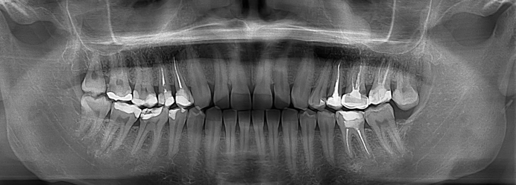
A panoramic dental X-ray is a non-invasive diagnostic test that is generally performed, by your dentist or oral surgeon, to diagnose medical conditions. It is performed so your dentist can obtain an initial evaluation of your maxillary sinuses, tooth position, bones and teeth. It can also be used to plan treatment for dentures, braces, extractions, or implants.
The panoramic X-ray is a two dimensional (2-D) X-ray that takes images of your mouth, in a single image, that includes your teeth, upper and lower jaw, and the surrounding structures and tissues. Even though your jaw is curved, the panoramic X-ray will produce a flat image of your curved mouth.
A panoramic X-ray can also provide information about dental and medical concerns, including:
- Cysts in the jaw bone
- Tumors in the jaw
- Oral cancer
- Impacted teeth
- Disorders of the jaw
- Sinusitis
- Advanced periodontal disease
There is no special preparation for this non-invasive diagnostic procedure. Please let your doctor know if there is a possibility that you are pregnant. You will be asked to wear a lead apron as a safety precaution to protect your body from exposure to radiation. You may be asked to remove any metal objects you are wearing so they don’t interfere with the X-ray images.
To prepare for the panoramic X-ray procedure, you will either stand or sit with your head positioned on a chin, forehead, and side rest. You will be provided with a bite blocker so your mouth is opened slightly and your teeth are aligned so a clear picture can be obtained. During the procedure, you will need to remain very still while an X-ray tube is rotated around your head, in a semicircle, from one side of your jaw to the other.